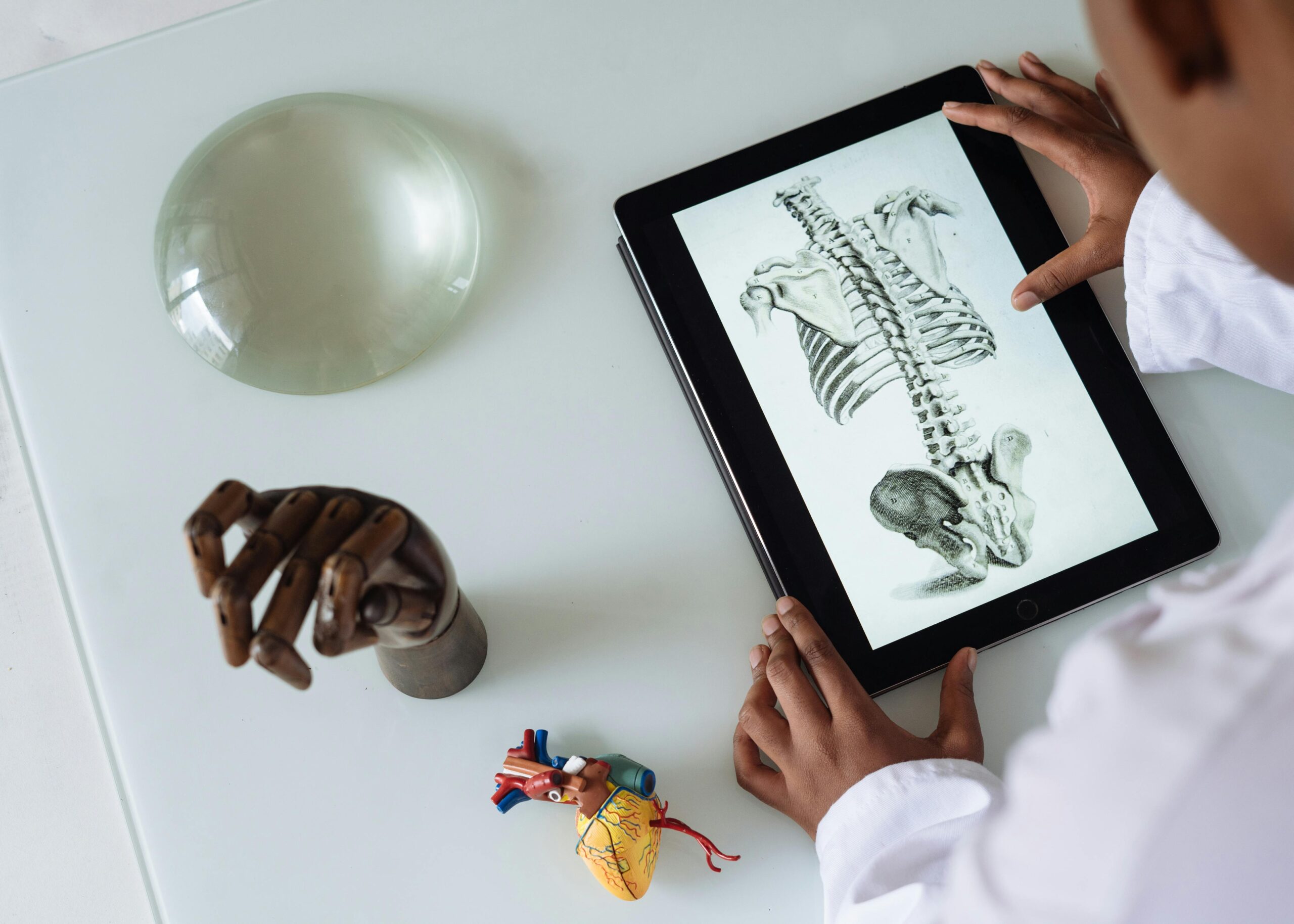
Clinical tissue regeneration is transforming modern medicine, offering hope where conventional treatments once reached their limits. This revolutionary field combines cutting-edge biotechnology with innovative therapeutic approaches.
From burn victims regaining functional skin to cardiac patients receiving regenerated heart tissue, the landscape of healing is undergoing a profound transformation. These advances represent not merely incremental improvements but fundamental shifts in how we approach tissue damage, disease, and the body’s innate capacity to heal itself.
🔬 The Dawn of Regenerative Medicine: Understanding the Foundation
Tissue regeneration harnesses the body’s natural healing mechanisms while amplifying them through scientific intervention. Unlike traditional medicine that often masks symptoms or replaces damaged tissue with artificial materials, regenerative approaches actually restore biological function at the cellular level.
The field encompasses multiple therapeutic modalities including stem cell therapy, tissue engineering, growth factor applications, and biomaterial scaffolds. Each approach targets specific aspects of the healing process, whether stimulating cell proliferation, guiding tissue organization, or creating conducive environments for regeneration.
What makes current regenerative medicine particularly exciting is the convergence of multiple scientific disciplines. Molecular biology, materials science, bioengineering, and clinical medicine now work in concert, creating solutions previously confined to science fiction.
Rebuilding Skin: The Burns Unit Breakthrough 🔥
One of the most visually dramatic applications of tissue regeneration involves severe burn treatment. Traditional approaches relied heavily on skin grafts harvested from the patient’s own body, limiting treatment options for extensive burns.
At Massachusetts General Hospital, researchers developed a spray-on skin technology that has transformed outcomes for burn patients. The technique involves harvesting a small skin sample, isolating stem cells and keratinocytes, and then suspending them in a solution that can be sprayed directly onto the wound.
In one remarkable case, a firefighter with third-degree burns covering 35% of his body received this treatment. Within weeks, new skin began forming across previously damaged areas. Six months post-treatment, the regenerated skin demonstrated normal elasticity, sensation, and appearance—outcomes that would have been impossible with conventional grafting alone.
The Science Behind Skin Regeneration
This approach works by providing the wound bed with concentrated populations of the cells responsible for skin formation. The spray delivery method ensures even distribution and allows treatment of irregular wound surfaces that would be difficult to cover with traditional grafts.
The regenerated tissue doesn’t just look like normal skin—it functions like it too. Sweat glands, hair follicles, and sensory nerves all develop within the new tissue, restoring not just coverage but genuine biological function.
Cardiac Tissue Regeneration: Healing the Heart ❤️
Heart disease remains the leading cause of death globally, with heart attacks causing permanent damage to cardiac muscle. Unlike skin or liver, the heart has extremely limited natural regenerative capacity. When cardiac tissue dies, it typically scars rather than regenerates.
Researchers at Stanford University pioneered an approach using cardiac progenitor cells—specialized cells with the potential to become heart muscle. In clinical trials, these cells were injected directly into damaged heart tissue following heart attacks.
One patient, a 58-year-old businessman who suffered a massive heart attack, participated in this trial. His ejection fraction—a measure of the heart’s pumping efficiency—had dropped to 25% (normal is 55-70%). Three months after receiving the cell therapy, his ejection fraction improved to 38%, and he reported significant improvements in stamina and quality of life.
Measuring Success in Cardiac Regeneration
The improvements weren’t merely subjective. Advanced imaging techniques revealed new blood vessel formation in previously damaged areas and improved contractility of the heart muscle. Biomarkers indicated that actual tissue regeneration, not just improved function of surviving tissue, was occurring.
While complete restoration of heart function remains elusive, these therapies represent a fundamental shift from managing heart disease to actually reversing damage.
Spinal Cord Injury: Reconnecting Neural Pathways 🧠
Perhaps no area of regenerative medicine captures the imagination quite like spinal cord injury treatment. The conventional medical wisdom held that spinal cord damage was permanent, with paralysis an inevitable consequence of severe injury.
A groundbreaking case from the University of California involved a 38-year-old woman paralyzed from the chest down following a car accident. Eighteen months post-injury, she received an experimental treatment combining neural stem cells with a supportive biomaterial scaffold.
The scaffold, made from a specialized hydrogel, was injected into the injury site where it formed a three-dimensional structure. This scaffold served as a bridge, providing physical support and biochemical signals that encouraged nerve fiber growth across the damaged area.
Remarkable Recovery and Functional Gains
Six months after treatment, the patient began experiencing tingling sensations in her legs—the first sensory feedback since her injury. By one year, she could move her toes voluntarily. At eighteen months post-treatment, she could stand with assistance and take several steps using a walker.
While not a complete cure, the functional improvements dramatically enhanced her independence and quality of life. More importantly, the case demonstrated that the “permanent” nature of spinal cord injury could be challenged with appropriate regenerative interventions.
Bone Regeneration: Engineering Structural Support 🦴
Orthopedic applications of tissue regeneration have advanced rapidly, addressing everything from non-healing fractures to bone cancer defects. Traditional approaches relied on bone grafts—either from the patient’s own body or cadaver donors—with significant limitations including limited supply, rejection risk, and incomplete integration.
At the Mayo Clinic, surgeons treated a 45-year-old construction worker whose tibia (shinbone) had failed to heal properly after a severe fracture. The bone gap measured nearly four centimeters—too large for natural healing.
The surgical team employed a tissue-engineered bone substitute combining a calcium phosphate scaffold with the patient’s own bone marrow cells, rich in mesenchymal stem cells capable of forming bone. The scaffold was precisely shaped to fit the defect using 3D printing technology based on CT scans of the patient’s leg.
Integration and Remodeling
Within three months, X-rays showed new bone formation bridging the gap. By six months, the engineered bone had integrated seamlessly with the patient’s existing bone structure. The scaffold gradually dissolved as it was replaced by natural bone through a process called remodeling.
The patient returned to work without limitations, bearing full weight on the previously damaged leg. Follow-up imaging two years later showed the regenerated bone was indistinguishable from natural bone in density and structure.
Cartilage Restoration: Addressing the Osteoarthritis Challenge 💪
Cartilage damage, whether from injury or degenerative disease, has long presented treatment challenges. Cartilage lacks blood supply, severely limiting its natural healing capacity. Once damaged, it typically deteriorates progressively, often leading to debilitating arthritis.
A 52-year-old former athlete with severe knee cartilage damage faced joint replacement surgery as his only option. Instead, he enrolled in a clinical trial at Hospital for Special Surgery in New York testing a novel cartilage regeneration approach.
The procedure involved arthroscopic surgery to prepare the damaged area, followed by injection of a gel containing chondrocytes (cartilage cells) derived from the patient’s own tissue, cultured and expanded in the laboratory.
Functional Outcomes and Long-Term Success
Within six weeks, the patient reported reduced pain and improved mobility. MRI scans at three months showed the formation of new cartilage-like tissue filling the previously damaged areas. At one year, the regenerated cartilage demonstrated mechanical properties similar to natural cartilage.
Five years post-procedure, the patient remains active, participating in cycling and swimming without pain. The regenerated cartilage has prevented the progressive deterioration typical of untreated cartilage damage, potentially eliminating or significantly delaying the need for joint replacement.
Liver Tissue Regeneration: Harnessing Natural Capacity 🏥
The liver possesses remarkable natural regenerative capacity, yet severe disease or acute injury can overwhelm this ability. End-stage liver disease traditionally required transplantation, with long waiting lists and significant risks.
Researchers at King’s College London developed a bioartificial liver support system combining liver cells grown on specialized scaffolds with dialysis-like technology. This system supports failing livers while simultaneously promoting regeneration of the patient’s own liver tissue.
A 34-year-old woman with acute liver failure due to medication toxicity had only days to live without transplant. She received the bioartificial liver support as a bridge therapy while awaiting transplant. Remarkably, after three weeks of support, her own liver began showing signs of recovery.
Recovery Without Transplantation
The support was gradually reduced as her liver function improved. Within two months, her liver had regenerated sufficiently that transplantation was no longer necessary. Five years later, she maintains normal liver function without requiring transplant.
This case exemplifies how regenerative approaches can work with the body’s natural healing capacity, providing temporary support that allows time for regeneration to occur.
Corneal Regeneration: Restoring the Gift of Sight 👁️
Corneal damage from injury, infection, or disease causes blindness in millions worldwide. While corneal transplantation is possible, donor tissue shortages limit access to treatment, particularly in developing nations.
A breakthrough approach developed in Sweden uses a biosynthetic cornea made from recombinant human collagen. Unlike donor corneas, these can be manufactured in large quantities and don’t require immunosuppression.
In a clinical trial, a 57-year-old man with corneal scarring that had left him legally blind received one of these biosynthetic corneas. The procedure involved removing the damaged cornea and suturing the biosynthetic replacement in its place.
Vision Restoration and Tissue Integration
Within weeks, the patient’s own cells began migrating into the biosynthetic material, gradually transforming it into living tissue. Six months post-surgery, his vision had improved from light perception only to 20/40—sufficient for reading and most daily activities.
The regenerated cornea remained clear and functional three years later, with the biosynthetic material having been completely replaced by the patient’s own cells and proteins, creating a natural, living cornea.
The Role of Stem Cells: Universal Building Blocks 🧬
Many regenerative successes rely on stem cells—cells with the remarkable ability to develop into many different cell types. Stem cells can be derived from various sources, each with distinct advantages.
Embryonic stem cells possess the greatest developmental potential but raise ethical concerns. Adult stem cells, found in bone marrow, fat tissue, and other locations, avoid these ethical issues but have more limited differentiation potential. Induced pluripotent stem cells (iPSCs), created by reprogramming adult cells, combine broad potential with ethical acceptability.
A particularly promising approach uses mesenchymal stem cells (MSCs) harvested from the patient’s own adipose (fat) tissue. These cells can differentiate into bone, cartilage, and other tissues, while also secreting growth factors that promote healing.
Biomaterials: The Scaffold for Regeneration 🏗️
Many tissue engineering approaches require biomaterial scaffolds—three-dimensional structures that provide physical support and biochemical cues guiding cell behavior and tissue formation.
Modern scaffolds are sophisticated structures designed to mimic natural tissue architecture. They must be biocompatible, avoiding immune rejection or toxic effects. They need appropriate mechanical properties, providing support while allowing cell migration. They should be biodegradable, dissolving as natural tissue replaces them.
Advanced manufacturing techniques including 3D printing allow creation of patient-specific scaffolds with precise geometry matching the defect being treated. Some scaffolds incorporate growth factors or genes that are gradually released, providing sustained regenerative signals.
Overcoming Challenges: The Path Forward 🚀
Despite remarkable successes, tissue regeneration faces significant challenges. Manufacturing complexity and costs currently limit widespread availability. Long-term outcomes remain uncertain for many approaches, requiring continued monitoring. Regulatory pathways for these advanced therapies continue evolving, sometimes creating delays in clinical availability.
Individual patient variability means treatments effective for some may fail for others, necessitating personalized approaches. The complexity of recreating functional tissue, particularly organs with intricate structures, remains beyond current capabilities.
Emerging Technologies and Future Directions
Next-generation approaches promise to address current limitations. Gene editing using CRISPR technology may enhance regenerative capacity of cells. Advanced bioreactors create controlled environments optimizing tissue development. Artificial intelligence accelerates identification of optimal cell types, scaffolds, and growth factor combinations.
Bioprinting technology is advancing toward printing functional organs. While still experimental, researchers have successfully printed simple tissues including skin, cartilage, and blood vessels. More complex organs remain challenging but may become feasible within decades.
Transforming Healthcare: The Broader Impact 🌟
The implications of successful tissue regeneration extend far beyond individual patients. These technologies could dramatically reduce the need for organ transplantation, eliminating waiting lists and the need for lifelong immunosuppression. They may transform chronic diseases from progressive conditions requiring management into potentially curable conditions.
Economic impacts could be substantial. While initial costs are high, regenerative treatments that provide lasting cures may prove more cost-effective than lifelong management of chronic conditions. Reduced disability and improved quality of life generate significant societal value beyond direct healthcare savings.
The convergence of regenerative medicine with other emerging technologies including artificial intelligence, nanotechnology, and advanced imaging creates synergistic possibilities. Personalized regenerative therapies tailored to individual patient genetics and disease characteristics represent the future of precision medicine.

Inspiring Hope Through Innovation 💡
The case studies presented here represent just a fraction of the remarkable advances occurring in clinical tissue regeneration. Each success story reflects years of painstaking research, clinical trials, and the courage of patients willing to try experimental treatments.
These are not distant future possibilities—they are happening now, transforming lives in clinics and hospitals worldwide. Patients who would have faced permanent disability or death now experience restored function and renewed hope.
As research continues and technologies mature, tissue regeneration will increasingly move from experimental to standard treatment. The question is no longer whether regenerative medicine will transform healthcare, but how quickly and completely this transformation will occur.
For patients currently facing conditions once considered untreatable, these advances offer genuine hope. For healthcare providers, they provide powerful new tools to heal rather than merely manage disease. For society, they promise reduced suffering and expanded human potential.
The revolution in healing through tissue regeneration has begun, and its impact on medicine and human health will only grow in the coming decades. These inspiring case studies illuminate not just what has been achieved, but what becomes possible when scientific innovation meets clinical need and human determination.
Toni Santos is a biomedical researcher and genomic engineer specializing in the study of CRISPR-based gene editing systems, precision genomic therapies, and the molecular architectures embedded in regenerative tissue design. Through an interdisciplinary and innovation-focused lens, Toni investigates how humanity has harnessed genetic code, cellular programming, and molecular assembly — across clinical applications, synthetic organisms, and engineered tissues. His work is grounded in a fascination with genomes not only as biological blueprints, but as editable substrates of therapeutic potential. From CRISPR therapeutic applications to synthetic cells and tissue scaffold engineering, Toni uncovers the molecular and design principles through which scientists reshape biology at the genomic and cellular level. With a background in genomic medicine and synthetic biology, Toni blends computational genomics with experimental bioengineering to reveal how gene editing can correct disease, reprogram function, and construct living tissue. As the creative mind behind Nuvtrox, Toni curates illustrated genomic pathways, synthetic biology prototypes, and engineering methodologies that advance the precision control of genes, cells, and regenerative materials. His work is a tribute to: The transformative potential of CRISPR Gene Editing Applications The clinical promise of Genomic Medicine and Precision Therapy The design innovations of Synthetic Biology Systems The regenerative architecture of Tissue Engineering and Cellular Scaffolds Whether you're a genomic clinician, synthetic biologist, or curious explorer of engineered biological systems, Toni invites you to explore the cutting edge of gene editing and tissue design — one base pair, one cell, one scaffold at a time.


